What is Histotripsy?
The term “histotripsy” is derived from two Greek root words. “Histo” means soft tissue, and “tripsy” means breakdown.
Histotripsy is a non-invasive process that uses sound waves to completely eliminate cancer tumors. Like radiation therapy, doctors point an ultrasound device at your tumor and “zap it.” But unlike radiotherapy, there is no cancer-causing radiation or heat involved, tumors can be destroyed in one treatment, there is minimal damage to surrounding tissue, a low rate of complications, faster recovery time, and it has been shown to activate immune cells to identify and target any remaining cancer cells in the body. [2]
The U.S. Food and Drug Administration has approved the use of sound waves to break down tumors — a technique called histotripsy, which was pioneered at the University of Michigan — in humans for liver treatment. [3]
“The future of medicine is the medicine of frequencies.” – Albert Einstein
The Science Behind Histotripsy: A Deeper Dive
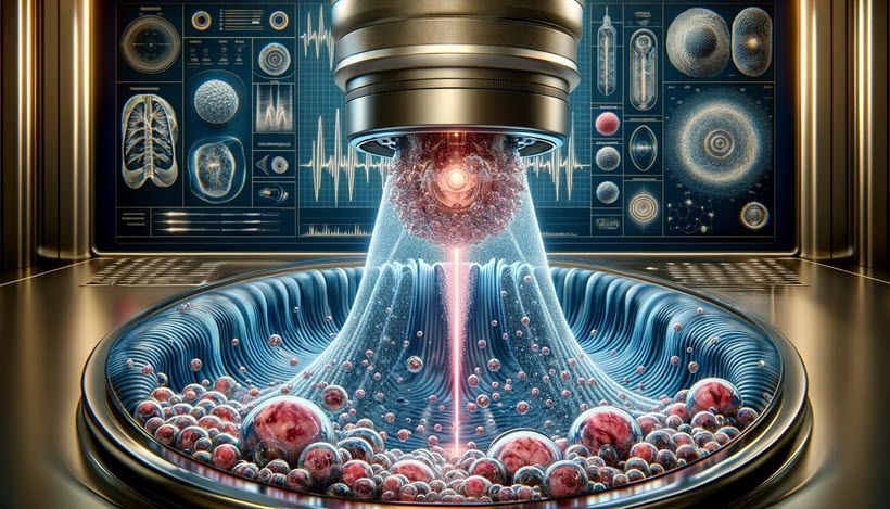
The Science: Histotripsy uses short ultrasound bursts (microseconds in length) with a low duty cycle (≤1%) to minimize heating, and higher peak pressure amplitudes to generate acoustic cavitation from endogenous gas in tissues. Acoustic cavitation is the generation, oscillation, and collapse of microbubbles activated by ultrasound. Very high ultrasound pressure causes inertial expansion and collapse of cavitation bubbles that impart localized intense strain that can fracture cells into an acellular debris. [1]
Translation: Histotripsy uses targeted high-intensity ultrasound, which causes tiny bubbles of gas inside cells to rapidly expand and collapse causing the cell to rupture, essentially blowing up cancer cells from the inside. The body then metabolizes and eliminates the cellular debris. [1]
Benefits of Histotripsy
Histotripsy offers many benefits for patients. Offering an additional treatment option to radiation, chemotherapy, and immunotherapy, histotripsy allows patients to avoid the physical toll and side effects often associated with these methods. Patients can also experience shorter recovery times and less discomfort.
In addition, histotripsy’s non-invasive approach means patients unable to tolerate more aggressive treatments have an effective alternative treatment option. After histotripsy treatment, most patients typically have no remaining viable tumor.
Histotripsy can precisely treat cancerous tissue, but it may not be suitable if tumors are near critical body structures. For this reason, histotripsy is less suitable as a treatment option for other forms of cancer such as breast, colorectal, pancreatic, lung, and others. [2]
What You Need to Know
- Histotripsy uses strong sound waves to target tumorous tissue in the liver.
- Because the ultrasound waves target only the tumorous tissue, they leave surrounding healthy tissue unharmed, which means a shorter recovery period for patients.
- Histotripsy doesn’t require any incisions to the body, minimizing potential complications and making recovery time much shorter than traditional surgery.
- The procedure poses fewer risks than those associated with traditional surgeries such as infection, scarring, pain and bleeding.
- Histotripsy is a procedure suited for patients for whom traditional surgical methods, chemotherapy or radiation pose increased risks or have been ineffective.
References
[1] Wark, Chris. 2024. “Histotripsy: Destroying Cancer Tumors with Sound Waves.” Chris Beat Cancer. December 16. https://www.chrisbeatcancer.com/histotripsy-destroying-cancer-tumors-with-sound-waves/.
[2] “Histotripsy.” 2025. cooperhealth.org. Accessed January 10. https://www.cooperhealth.org/services/histotripsy.
[3] Jim Lynch College of Engineering. 2025. “FDA Approves Histotripsy for Liver Treatment in Humans.” The University Record. Accessed January 10. https://record.umich.edu/articles/fda-approves-histotripsy-for-liver-treatment-in-humans/.
Videos
Additional Reading
“Histotripsy.” 2025. UChicago Medicine. Accessed January 10. https://www.uchicagomedicine.org/cancer/types-treatments/histotripsy.
Histotripsy is an innovative, non-invasive treatment for liver tumors that uses a robotic machine to target and destroy cancer tissue with ultra-precise sound waves. It’s a powerful tool for doctors to treat certain tumors without any needles, radiation or surgery, even allowing most patients to go home the same day.
“Histotripsy for Liver Tumors.” 2024. Johns Hopkins Medicine. December 23. https://www.hopkinsmedicine.org/health/treatment-tests-and-therapies/histotripsy-for-liver-tumors.
Histotripsy is an interventional radiology procedure that uses focused ultrasound waves to treat liver tumors, both cancerous and benign. The treatment is delivered without needing to cut into the body.
“Research.” 2023. Histotripsy Group. August 17. https://histotripsy.umich.edu/research/.
Histotripsy was invented and developed in our lab. Histotripsy uses microsecond-length ultrasound pulses to break down the target tissue into acellular debris mechanically. We studied the mechanisms underlying histotripsy. As histotripsy uses microsecond-length pulses at extremely high pressure, specialized instrumentation is required. We have designed and built specialized instrumentation, including ultrasound transducers and driving electronics, to enable histotripsy. We have also made many technical advancements, including aberration correction and treatment monitoring, to ensure the safety and efficacy of histotripsy. Most recently, we have focused on developing histotripsy for various clinical applications, including the treatment of cancer, brain diseases, and cardiovascular diseases. Our research has led to the formation of HistoSonics (www.histosonics.com), a company based in Minneapolis and Ann Arbor that is commercializing histotripsy for oncological applications.
Wark, Chris. “chrisbeatcancer.” 2025. Instagram. Accessed January 10. https://www.instagram.com/reel/DDwzVTgSdgE/?igsh=aGxhN293OXNiZTF4.
Like radiation therapy, doctors point a device at your tumor and “zap it.” But unlike radiotherapy, there is no cancer-causing radiation used or heat involved. Tumors can be destroyed in one 2-hour histotripsy treatment. There is minimal damage to surrounding tissue, a low rate of complications, and a faster recovery time. Histotripsy has even been shown to activate immune cells to identify and target remaining cancer cells in the body.
Xu, Zhen, Timothy L Hall, Eli Vlaisavljevich, and Fred T Lee. 2025. “Histotripsy: The First Noninvasive, Non-Ionizing, Non-Thermal Ablation Technique Based on Ultrasound.” International Journal of Hyperthermia : The Official Journal of European Society for Hyperthermic Oncology, North American Hyperthermia Group. U.S. National Library of Medicine. Accessed January 10. https://pmc.ncbi.nlm.nih.gov/articles/PMC9404673/.
Histotripsy is the first noninvasive, non-ionizing, and non-thermal ablation technology guided by real-time imaging. Using focused ultrasound delivered from outside the body, histotripsy mechanically destroys tissue through cavitation, rendering the target into acellular debris. The body absorbs the material in the histotripsy ablation zone within 1–2 months, leaving a minimal remnant scar. Histotripsy has also been shown to stimulate an immune response and induce abscopal effects in animal models, which may have positive implications for future cancer treatment. Histotripsy has been investigated for a wide range of applications in preclinical studies, including treating cancer, neurological diseases, and cardiovascular diseases. Three human clinical trials have been undertaken using histotripsy for the treatment of benign prostatic hyperplasia, liver cancer, and calcified valve stenosis. This review provides a comprehensive overview of histotripsy, covering the origin, mechanism, bioeffects, parameters, instruments, and the latest preclinical and human studies results.
The featured image on this page is from the Vibramar website.
